41 human heart diagram and labels
Histology guide: Definition and slides - Kenhub At a histological level, both the heart and blood vessels consist of three layers: Endothelial layer - epithelial tissue formed by simple squamous (endothelial) cells. In the heart, this layer is referred to as endocardium. Muscular layer - smooth muscle in the blood vessels, cardiac muscle (myocardium) in the heart. What Exactly Is Human Anatomy? - Health Pages The regions are named below and the corresponding regions are labeled 1-9. Abdominal Regions Right (1) and left (3) hypochondriac regions - on either side of the epigastric region. Contains the diaphragm, some of the kidneys, right side of the liver, the spleen and part of the pancreas.
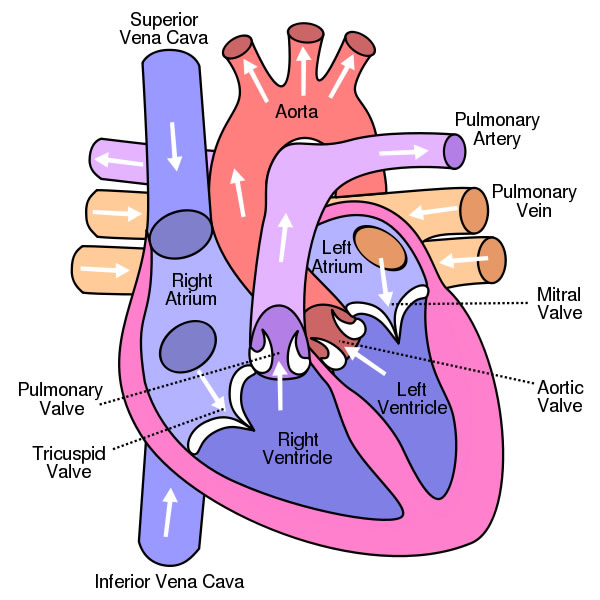
Heart Anatomy The Worksheet Of Label the heart as a box on the board together - use the animation on the Powerpoint to illustrate the cardiac cycle once labelling is complete Sketch the heart cross-section and identify the following components: ventricles, atriums, pericardium, Map of the Human Heart superior vena cava 6 . superior vena cava 6

Human heart diagram and labels
CB1R-stabilized NLRP3 inflammasome drives antipsychotics cardiotoxicity ... h The simulated interaction diagram of human CB1R peptide (amino acids 177-189) with human proteins NLRP3 (up, pink cartoon), CASP1 (middle, yellow cartoon), or GSDMD (down, gray cartoon) in left ... Path of Blood Through the Heart - New Health Advisor Here are the basic parts of the heart: 1. Right Atrium The heart can be divided into right and left halves, as well as into the upper and lower chambers. There are two upper chambers called atria and two lower chambers called ventricles. On each side of the heart, there are one atrium and one ventricle. Positions and Functions of the Four Brain Lobes - MD-Health.com The human brain contains the frontal, occipital, temporal, and parietal lobes. Learn how the brain lobes function to support our thoughts and reactions. The human brain is the most complex organ in the body. Composed of 50 to 100 billion neurons, the human brain remains one of the world's greatest unsolved mysteries. Here we will take a closer ...
Human heart diagram and labels. [Life Processes] Similarities & difference between Aerobic & Anaerobic Aerobic respiration occurs in the presence of oxygen. 1. Anaerobic respiration occurs in the absence of oxygen. 2. Complete oxidation of glucose takes place to form CO 2 and H 2 O. 2. Incomplete oxidation of glucose takes place to form alcohol / lactic acid and CO 2. 3. It occurs in plant and animal cells. Types of tissue: Structure and function - Kenhub There are four basic tissue types defined by their morphology and function: epithelial tissue, connective tissue, muscle tissue, and nervous tissue. Epithelial tissue creates protective boundaries and is involved in the diffusion of ions and molecules. Connective tissue underlies and supports other tissue types. Personalized Medicine | National Institutes of Health (NIH) The Human Genome Project and thousands of follow-on studies are helping scientists to develop gene-targeted treatments. A poignant example is the case of a woman with lung cancer that had spread to her brain. Diagnosed in 2002, this 44-year-old—a vegetarian who had never smoked—underwent various therapies to stave off what seemed inevitable. Mechanism of Respiration in Human Beings - Embibe The human respiratory system consists of a group of organs that help to breathe. Lungs are the primary organs of the respiratory system that comprises about \(600-700\) million alveoli (air sacs). ... Because of this, the blood cannot pick up the oxygen from the alveoli and causes damage to the heart and kidneys. Important Facts. Respiration ...
Trunk Region (Torso) < Regional Anatomy << Human Anatomy <<< Body ... Trunk (Torso) In our body, the Trunk Region (Torso), an anatomical term for the central part of the body, is a combination of both the thoracic region (chest), including the mammary region (breasts), and the abdomen region (belly region) including the naval (umbilicus region), coxal region, and pubic region. In our body, the Trunk Region (Torso) as an anatomical term for the central part of ... Circulatory system - Wikipedia The blood circulatory system, is a system of organs that includes the heart, blood vessels, and blood which is circulated throughout the entire body of a human or other vertebrate. [1] [2] It includes the cardiovascular system, or vascular system, that consists of the heart and blood vessels (from Greek kardia meaning heart, and from Latin ... Diagram of Human Heart and Blood Circulation in It Exterior of the Human Heart A heart diagram labeled will provide plenty of information about the structure of your heart, including the wall of your heart. The wall of the heart has three different layers, such as the Myocardium, the Epicardium, and the Endocardium. Here's more about these three layers. Epicardium Cerebral cortex: Structure and functions | Kenhub It contains three to six layers and is found in the insula, cingulate and parahippocampal gyri. The neocortex, as its name suggests, is the most recent cortical region and makes up to 90% of the human cortex. It includes all of the lobes of the cortex except the limbic lobe and consists of six layers of cells or laminae.
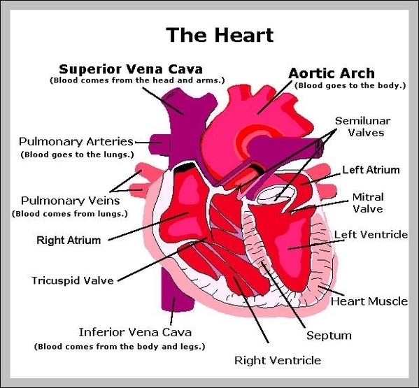
Charts of Normal Resting and Exercising Heart Rate The heart is an organ located just behind and slightly to the left of the breastbone, and pumps blood through a network of veins and arteries known as the circulatory system. The right atrium is sent blood from the veins, and delivers it to the right ventricle. It's then pumped into the lungs where it is oxygenated. Know Where Your Heart Is and How to Identify Heart Pain The heart is enclosed in the pericardium which is a double layer. This pericardium is attached to the diaphragm, spinal column and other parts via strong ligaments. Inside, heart is hollow and divided into 4 chambers: the upper 2 chambers are called left and right atria whereas the lower ones are called the left and right ventricles. Circulatory System Diagram - New Health Advisor Coronary circuit mainly consists of cardiac veins including anterior cardiac vein, small vein, middle vein and great (large) cardiac vein. There are different types of circulatory system diagrams; some have labels while others don't. The color blue stands for deoxygenated blood while red stands for blood which is oxygenated. Structure of Cell: Definition, Types, Diagram, Functions - Embibe Structure of Cell: Cell is the basic functional unit that makes up all living organisms.All organisms, including ourselves, start life as a single cell called the egg. Cells are small microscopic units that perform all essential functions of life and are capable of independent existence.
An easy diagram of HUMAN HEART | Homework Help | myCBSEguide
Lymphatic System: Lymphatic Functions, Diagram at Embibe They are formed of an outer coat of fibrous tissue, a middle coat of muscular tissue and an inner lining of endothelial cells. They have numerous valves. They are present in all tissues except the central nervous system and cornea. Structure ofLymphatic vessel D. Lymphatic Nodes

Heart Diagram Quiz
Nervous system: Structure, function and diagram | Kenhub The nervous system is a network of neurons whose main feature is to generate, modulate and transmit information between all the different parts of the human body. This property enables many important functions of the nervous system, such as regulation of vital body functions ( heartbeat, breathing, digestion), sensation and body movements.

The Heart Diagrams Labeled and Unlabeled
Free Human Anatomy Coloring Pages for Students The Human Skeleton Coloring Page - This anatomy coloring page involves labeling too - so you can visualize each part of the human skeleton. Skeletal System Worksheets - Our body's framework is made up of our skeletal system. These worksheets and printables will help your kids learn more about the bones in our body and how they work together.

Kids' Health - Topics - Your heart | Heart for kids, Heart diagram, Heart lesson
Eyeball: Structure and function | Kenhub Eyeball (Bulbus oculi) The eye is a highly specialized sensory organ located within the bony orbit.The main function of the eye is to detect the visual stimuli (photoreception) and to convey the gathered information to the brain via the optic nerve (CN II).In the brain, the information from the eye is processed and ultimately translated into an image.
Free Unlabelled Diagram Of The Heart, Download Free Unlabelled Diagram Of The Heart png images ...
Learn about Stages of Embryonic Development, Process - Embibe Learn about Stages of Embryonic Development in the human. In Biology, Embryonic Development is also called Embryogenesis. Learn more @Embibe. ... Embryo Development in Humans Diagram. Fig: Embryo and Fetal Development in Humans ... The heart is the first organ to start functioning during Embryonic Development. The heartbeat of an Embryo can be ...
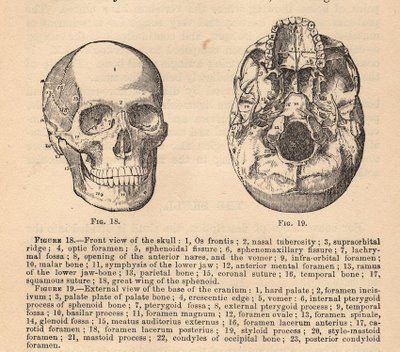
Vintage Graphic - Anatomy - Skull Diagram - The Graphics Fairy
Heart - Life processes - Class Notes Heart (1) It is a muscular organ, big as our fist, reddish brown in colour, situated between the 2 lungs in middle of thoracic cavity, surrounded by 2 layered sac. (2) It has different chambers to prevent the oxygen rich blood from mixing with blood containing carbon dioxide. (3) Heart is divided by septa into 2 halves i.e. the right and left.

HEALTH CARE ( The topic you are looking for...): January 2010
Parts of Human Eye and Their Functions - MD-Health.com The iris is the area of the eye that contains the pigment which gives the eye its color. This area surrounds the pupil, and uses the dilator pupillae muscles to widen or close the pupil. This allows the eye to take in more or less light depending on how bright it is around you. If it is too bright, the iris will shrink the pupil so that they ...

Labelled Diagram of the Human Heart - Cancer / Asbestos Cancer | Diagram of Heart | Mesothelioma >>
Human Circulatory System: Definition, Organs and Functions The human heart consists of four chambers - two ventricles and two auricles. Organs or Parts of Circulatory System The main parts of the human circulatory system include organs like blood vessels, the lymphatic system and the heart, and a fluid connective tissue called the blood. Let us now learn about the organs of the circulatory system below:

Human Heart Diagram
Parts and Components of Human Ear and Their Functions In addition to helping the body take in auditory messages, the ear helps to maintain a proper head position. The fluid in the ear also helps the body maintain a sense of balance so the body can maintain proper posture and coordination. There are three major parts of the ear, the outer, middle and inner ear. Each contains several parts that are ...
Human Heart Diagram - Human Body Pictures - Science for Kids
How would you test the presence of starch in leaves? Answer: When the leaves are boiled in alcohol, they lose chlorophyll, which is a green colored pigment, and become colorless. The end product after photosynthesis is starch. Starch is stored in the leaves. It can be tested for by adding iodine solution which gives a bluish black colour indicating the presence of starch in the leaf.
heart parts – Graph Diagram
Positions and Functions of the Four Brain Lobes - MD-Health.com The human brain contains the frontal, occipital, temporal, and parietal lobes. Learn how the brain lobes function to support our thoughts and reactions. The human brain is the most complex organ in the body. Composed of 50 to 100 billion neurons, the human brain remains one of the world's greatest unsolved mysteries. Here we will take a closer ...

Heart Diagram Worksheet Blank Human Heart Worksheets Label Heart Diagram Worksheet Awesome in ...
Path of Blood Through the Heart - New Health Advisor Here are the basic parts of the heart: 1. Right Atrium The heart can be divided into right and left halves, as well as into the upper and lower chambers. There are two upper chambers called atria and two lower chambers called ventricles. On each side of the heart, there are one atrium and one ventricle.

Anatomy of the Heart (Latin)
CB1R-stabilized NLRP3 inflammasome drives antipsychotics cardiotoxicity ... h The simulated interaction diagram of human CB1R peptide (amino acids 177-189) with human proteins NLRP3 (up, pink cartoon), CASP1 (middle, yellow cartoon), or GSDMD (down, gray cartoon) in left ...

Well Labelled Diagram Of Human Digestive System - Made By Creative Label
13+ Heart Diagram Templates – Sample, Example, Format Download | Free & Premium Templates

Label the Heart Quiz

282 best images about Diagramatically Speaking on Pinterest | Respiratory system, Neurons and ...
Post a Comment for "41 human heart diagram and labels"