40 eye diagram and labels
Circulatory System Diagram | New Health Advisor There are different types of circulatory system diagrams; some have labels while others don't. The color blue stands for deoxygenated blood while red stands for blood which is oxygenated. Below you'll see diagram specified to the heart, as well as circulatory system diagram of the whole body: How Does the Human Circulatory System Work? 1. Heart Object Labeling | Know Your Meme Object Labeling refers to the practice of creating image macros in which subjects of a specific image are labeled to create a humorous interpretation of the picture. Similar to exploitables, Object Labeling involves changing a picture in various ways to make different jokes. The practice began seeing use in meme making in the mid 2010s, as several meme formats from the time employed object ...
WHMIS 2015 - Pictograms : OSH Answers - Canadian Centre for ... The exclamation mark pictogram is used for the following classes and categories: Acute toxicity - Oral, Dermal, Inhalation (Category 4) Skin corrosion/irritation - Skin irritation (Category 2) Serious eye damage/eye irritation - Eye irritation (Category 2 and 2A) Respiratory or skin sensitization - Skin sensitizer (Category 1, 1A and 1B)

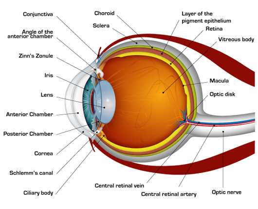
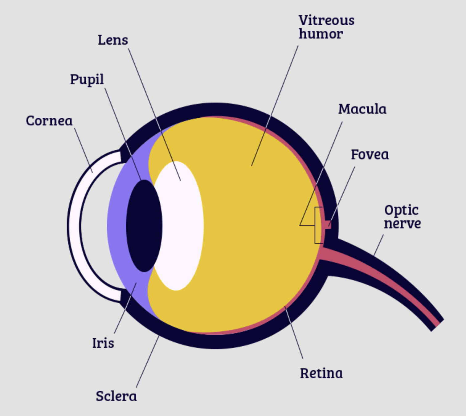
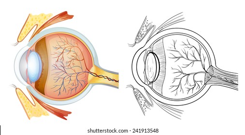
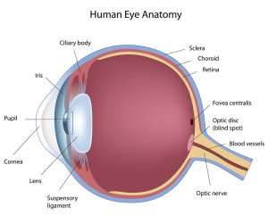
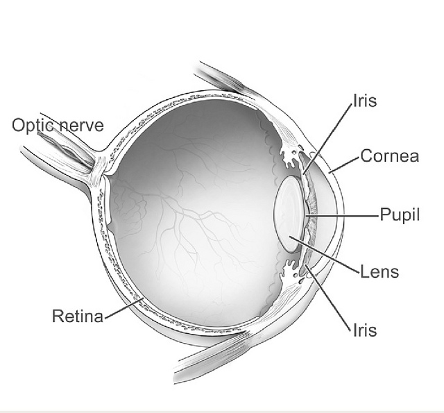
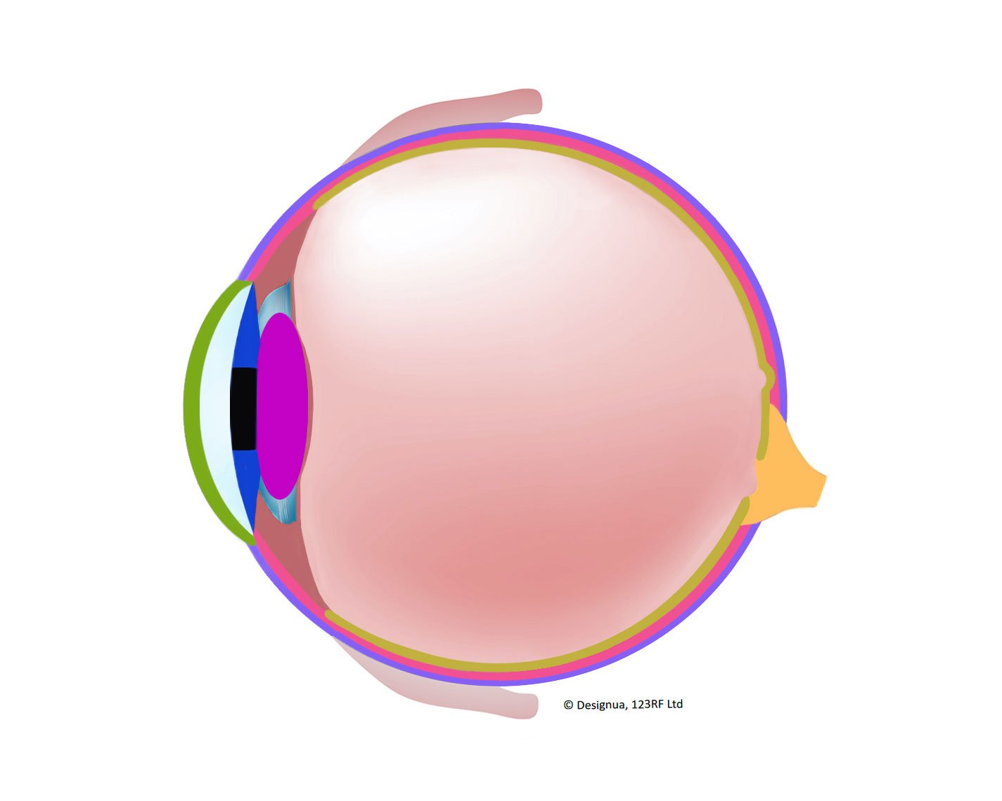
Eye diagram and labels
30 Amazing Facts About Human Eyes For Kids, With Diagrams - MomJunction Our eyes capture light and send the data to the brain for forming images. Red eyes in photographs appear due to the light from the flash bouncing off our capillaries in the eyes. If the human eye were a digital camera, it would have a resolution of 576 megapixels. The iPhone 12 has 12 megapixels. Quiz: Label The Parts Of The Eye - ProProfs Quiz Quiz: Label The Parts Of The Eye. Do you know the anatomy of the human eye very well? Can you label the parts of the eye in the quiz below? Give it a try and evaluate yourself. The eye has many important parts, each with different functions, including the cornea, pupil, sclera, and many more. Can you tell where these parts are located and what ... Microscope, Microscope Parts, Labeled Diagram, and Functions The Iris Diaphragm is located above the condenser lens and below the microscope stage. The different sized holes in the diaphragm helps to vary the size of the cone and intensity of light that is projected upward into the slide. However, there is no set rule regarding which setting to use for a particular power.
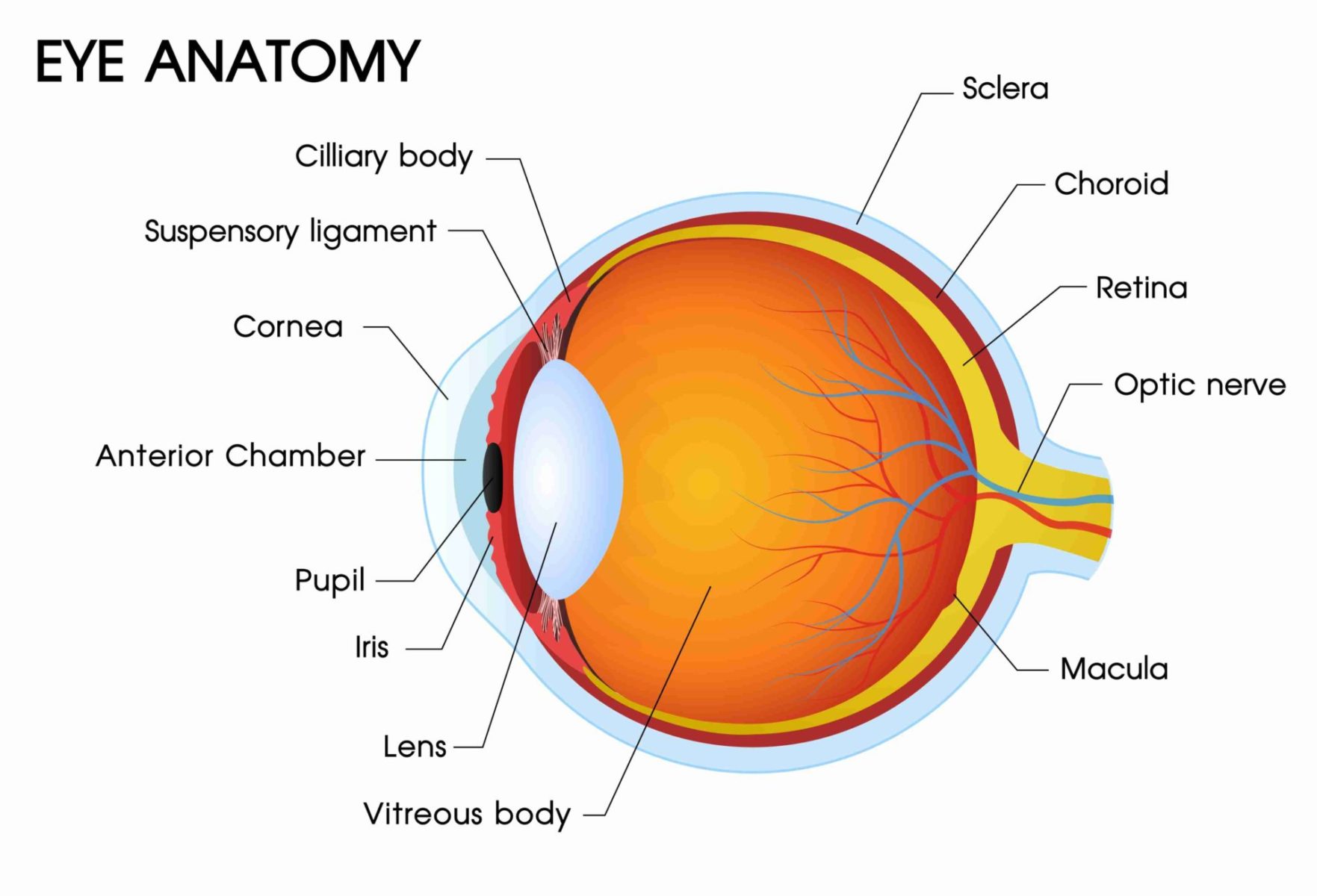
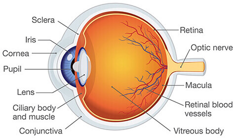
Eye diagram and labels. What To Know When You Order Online From Vision Direct Vision Direct offers next-day delivery to its U.K. customers. U.S. customers can get contacts from Vision Direct in about a week (five to seven business days). Orders placed over the weekend will ship out on Monday. Shipping to the United States on orders £59 and over (about $65 U.S. dollars) costs £9.98 (about $11 U.S. dollars). Anatomy of the eye: Quizzes and diagrams | Kenhub One of our favorite ways to get to grips with all of the parts of the eye is by utilizing labeled diagrams. On a diagram of the eye, we can see all of the relevant structures together on one image. This helps us to understand how each one is situated and related to the other. Labeled diagram of the eye Ear Diagram Quiz - ProProfs Quiz Take this ear diagram quiz, and see how well you understand the ear. Here, we have given a diagram where the labeling is done with the numbers. You just need to tell the name of the part labeled with the number asked in the question. It is a kind of practice test for you if you are preparing for an exam. Start the quiz, and see how much you score. The Most Dangerous Games: O Jogo das Tres Formas, Or The Three Cups Game O Jogo das Três Formas (which is sometimes written as O Jogo das 3 Formas) doesn't seem to have circulated very widely. I've seen it only in a few places — a now-defunct horror Tumblr, a now-defunct horror Facebook page, and a horror-centric forum called Terra Proibida, or Cursed Land. Dated Nov. 13, 2012, the Terra Proibida version is the oldest one of the three; however, the Facebook ...
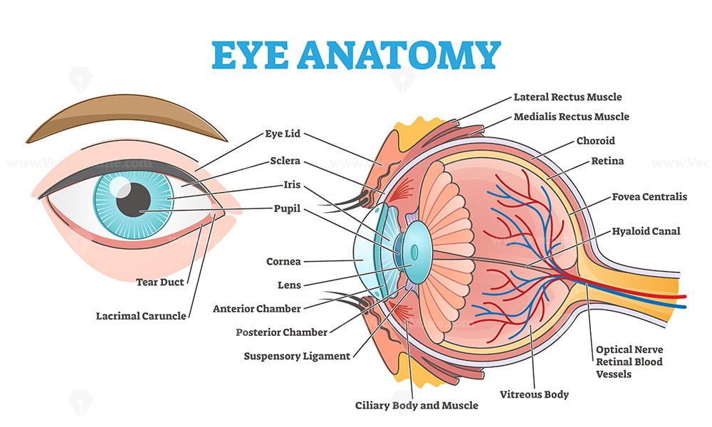
What's An Eye Tooth? | Colgate® Eye teeth have remarkably long roots and prominent crowns - all the better to help support the structure of your mouth. Eyeteeth also play the following roles in your mouth: Primal Role: With their more pointed and tapered shape, your cuspids help you grip and pierce through food easily as you bite. Leadership Role: And though they're not ... Brigitte Zimmer a typical animal cell diagram labeled black and white anatomical heart drawing with labels animal and plant cell diagram for class 9 ... draw a human eye and label it draw a picture of a plant cell in cytokinesis draw human digestive system drawing easy with label Eye Anatomy: 16 Parts of the Eye & Their Functions - Vision Center The following are parts of the human eyes and their functions: 1. Conjunctiva The conjunctiva is the membrane covering the sclera (white portion of your eye). The conjunctiva also covers the interior of your eyelids. Conjunctivitis, often known as pink eye, occurs when this thin membrane becomes inflamed or swollen. Eye Anatomy | Blood supply - Orbit - Extraocular muscles - Geeky Medics The eye can be divided into fibrous, vascular and inner layers. Fibrous layer The fibrous layer is the outermost layer and consists of the cornea and sclera, which are continuous with one another. The cornea is located in the centre of the anterior aspect of the eye and is transparent The sclera, which covers the rest of the eye, is white
Skin Layers: Structure, Function, Anatomy, and More - Verywell Health Its thickness depends on where it is on the body. It's thinnest on the eyelids (roughly half a millimeter) and thickest on your palms and soles (1.5 millimeters). The epidermis is made up of five layers. Stratum Corneum The stratum corneum is the top layer of the epidermis. Its jobs are to: Helps your skin retain moisture Bipolar Neurons - Structure and Functions | GetBodySmart 2. 3. Bipolar neurons are found in the retina of the eye, roof of the nasal cavity, and inner ear. They are always sensory and carry information about vision, olfaction, equilibrium, and hearing. In the eye, bipolar neurons form the middle layer of the retina. 1. 2. Here they conduct impulses from photoreceptors (rods and cones) to ganglion cells. human ear | Structure, Function, & Parts | Britannica The outer ear consists of the visible portion called the auricle, or pinna, which projects from the side of the head, and the short external auditory canal, the inner end of which is closed by the tympanic membrane, commonly called the eardrum. The function of the outer ear is to collect sound waves and guide them to the tympanic membrane. Teeth Numbers and Names - Human Teeth Chart - Dayo Dental Teeth numbers 14 and 15 are your upper left molars. If you are getting cosmetic dentistry using veneers, you usually want to enhance the most visible part, teeth numbers 6 - 11 on the upper and 22 - 26 on the lower. For movie fans, vampires can extend their eye teeth (canines): 6, 11, 22 and 27. Teeth Numbers and Names
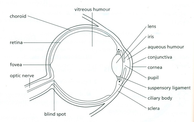
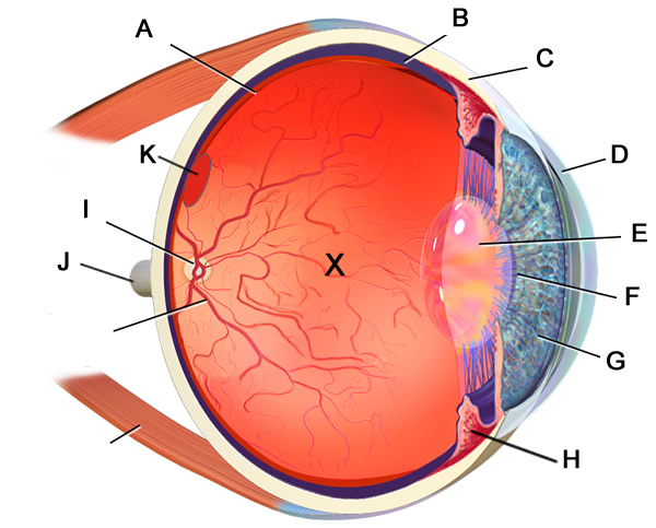
IGCSE Label the Eye Diagram | Quizlet
General Anatomy and Physiology of a Human: TEAS - Registered nursing Anatomy: The study of the parts and structures of the human body. Physiology: The study of the functions of the human body. Gross anatomy: The study of the parts and structures of the human body that can be seen with the naked eye and without the use of a microscope. Microscopic anatomy: The study of the parts and structures of the human body ...

Labeled Eye Diagram | Eye anatomy diagram, Eye anatomy ...
Ocumetics Bionic Eye Lens Updates | Disabled World Human Eye Diagram This diagram shows a labeled basic structure of the human eye with the three main layers - image courtesy of Wikimedia Commons, Artwork by Holly Fischer. Bionic Lens Updates May 18, 2022 : Ocumetics Update on the Status of Preclinical Studies for the Bionic Lens
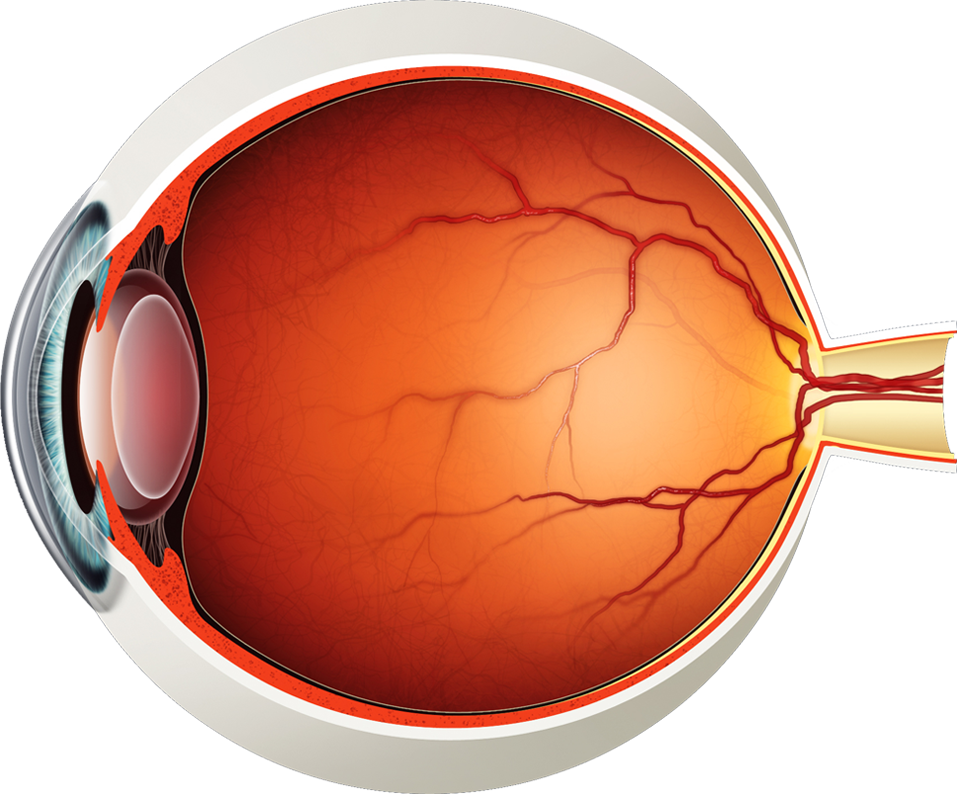
Diagram of the Eye - Lions Eye Institute
90 Car Dashboard Symbols, Warning Lights & Indicators - Mechanic Base Key Not in Vehicle Indicator. A Key Not in Vehicle symbol indicates the car's immobilizer system can't reach or recognize your car key. 77. Immobilizer Indicator. An immobilizer symbol means that it can't reach or recognize your car key, or an issue with the immobilizer system. 78. Ignition Switch Warning.

Structure Of Human Eye Without Label Transparent PNG ...
WHMIS 2015 - Labels : OSH Answers - Canadian Centre for Occupational ... Labels will require the following: the pictogram, signal word, and hazard statement are to be grouped together, to be clearly and prominently displayed on the container, to be easy to read (e.g., you can see it easily without using any item except corrective glasses), and to be in contrast with other information on the product or container.

Label Parts of the Human Eye
Parts and Components of Human Ear and Their Functions In addition to helping the body take in auditory messages, the ear helps to maintain a proper head position. The fluid in the ear also helps the body maintain a sense of balance so the body can maintain proper posture and coordination. There are three major parts of the ear, the outer, middle and inner ear. Each contains several parts that are ...

The human eye diagram (COMS). | Download Scientific Diagram
Eye Prescriptions (Meaning of SPH, CYL, AXIS) - Vision Center Axis refers to the lens meridian with no cylindrical power to correct astigmatism. The axis is labeled as a number from 1 to 180. If a prescription includes cylinder power, it must be followed by an axis. The axis value is often preceded by an "x" when written in freehand.

Human Eye Anatomy - Parts of the Eye Explained | Eye anatomy ...
Parts of the brain: Learn with diagrams and quizzes | Kenhub First up, have a look at the labeled brain structures on the image below. Try to memorize the name and location of each structure, then proceed to test yourself with the blank brain diagram provided below. Labeled diagram showing the main parts of the brain Blank brain diagram (free download!)

Sensory Structures | BioNinja
Anatomy of lower extremity - e-Anatomy - IMAIOS A diagram shows the various inguinal lymph nodes (lymphatic ganglia). The chapter on the innervation of the lower limb presents diagrams of the lumbosacral plexus and its main nerve branches for the lower limb (lateral cutaneous nerve of the thigh, femoral nerve, sciatic nerve and posterior cutaneous nerve of the thigh and obturator nerve).
How the Human Eye Works | Cornea Layers/Role | Light Rays
Heart: illustrated anatomy - e-Anatomy - IMAIOS This interactive atlas of human heart anatomy is based on medical illustrations and cadaver photography. The user can show or hide the anatomical labels which provide a useful tool to create illustrations perfectly adapted for teaching. Anatomy of the heart: anatomical illustrations and structures, 3D model and photographs of dissection.
About the Eye | National Eye Institute
Diagram of Human Heart and Blood Circulation in It Every heart diagram labeledwill clearly show these valves. These valves allow blood flow in one direction only. Different valves perform different functions. Tricuspid valve is located between the right ventricle of your heart and the right atrium, and allows the blood to move from the right atrium to the right ventricle.
13,140 Eye diagram Images, Stock Photos & Vectors | Shutterstock
Foot Anatomy and Common Foot Problems - Verywell Health Plus, the foot must be flexible to adapt to uneven surfaces and remain stable. Common foot problems include plantar fasciitis, bunions, flat feet, heel spurs, mallet toe, metatarsalgia, claw toe, and Morton's neuroma. This article provides an overview of foot anatomy and foot problems that come from overuse, injury, and normal wear and tear of ...
The Eye Anatomy | Vision Pro Optical | Eye Care You Can Trust
Microscope, Microscope Parts, Labeled Diagram, and Functions The Iris Diaphragm is located above the condenser lens and below the microscope stage. The different sized holes in the diaphragm helps to vary the size of the cone and intensity of light that is projected upward into the slide. However, there is no set rule regarding which setting to use for a particular power.
Eye Anatomy: 9 Main Parts of the Eye & How We See | Specialty ...
Quiz: Label The Parts Of The Eye - ProProfs Quiz Quiz: Label The Parts Of The Eye. Do you know the anatomy of the human eye very well? Can you label the parts of the eye in the quiz below? Give it a try and evaluate yourself. The eye has many important parts, each with different functions, including the cornea, pupil, sclera, and many more. Can you tell where these parts are located and what ...
The eye, rods and cones - Biology Notes for IGCSE 2014
30 Amazing Facts About Human Eyes For Kids, With Diagrams - MomJunction Our eyes capture light and send the data to the brain for forming images. Red eyes in photographs appear due to the light from the flash bouncing off our capillaries in the eyes. If the human eye were a digital camera, it would have a resolution of 576 megapixels. The iPhone 12 has 12 megapixels.
Eye anatomy with labeled structure scheme for human optic outline diagram

Cow's Eye Dissection - Eye diagram
File:Human eye diagram-sagittal view-NEI.jpg - Wikimedia Commons

Eye diagram by Firkin | Human eye diagram, Diagram of the eye ...
Label the Eye

Diagram human eye anatomy with label Royalty Free Vector

Eye pattern - Wikipedia
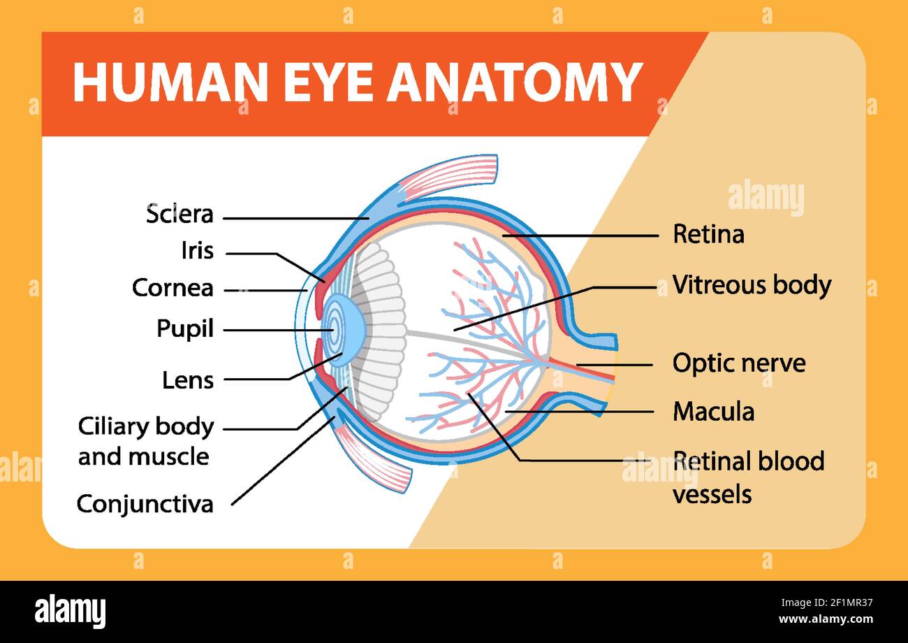
Diagram of human eye anatomy with label illustration Stock ...

Eye labeling Diagram | Quizlet
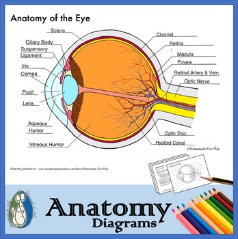
Anatomy of the Eye Diagrams for Coloring/Labeling, with ...
Label Eye Printout - EnchantedLearning.com
Eye Anatomy Diagram - EnchantedLearning.com
What Does the Eye Look Like? – Diagram of the Eye | Harvard ...

Eye anatomy and function

a Draw a labelled diagram of the human eye. Label the ...
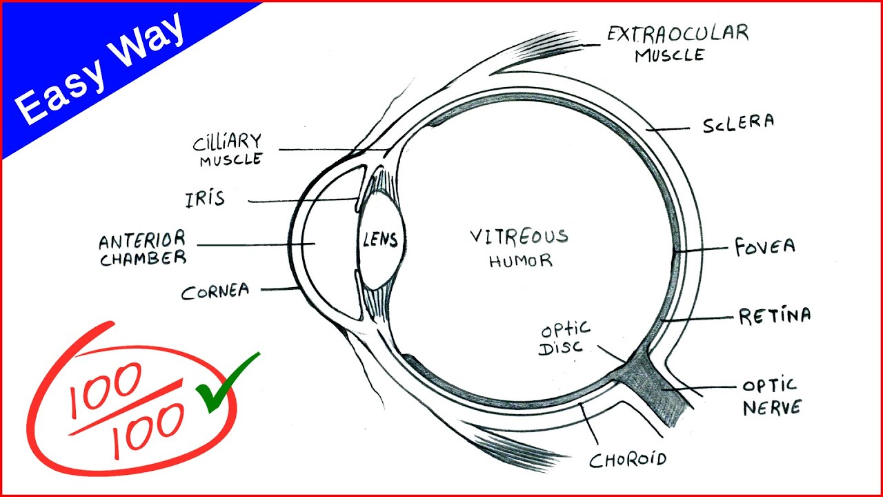
Eye Diagram drawing CBSE || easy way || draw Human eye anatomy - Step by step for beginners

Diagram human eye anatomy with label Royalty Free Vector
Neuroscience for Kids - Fill In #3

Label the Eye Quiz

Solved: Label the diagram. Refer to Figure 43-18 to check ...

Human Eye Diagram High-Res Vector Graphic - Getty Images

Eye Anatomy (labelled), illustration - Stock Image - C043 ...
/GettyImages-695204442-b9320f82932c49bcac765167b95f4af6.jpg)
Structure and Function of the Human Eye
Labelling the eye — Science Learning Hub

A schematic diagram of a horizontal section through an eye ...
Eye Doctor Manchester | Eye Anatomy Bedford | Medical Eye Center
Post a Comment for "40 eye diagram and labels"